
Myocardial Infarction (Heart Attack) in 3D | STEMI vs NSTEMI & MONA Protocol
Comprehensive Clinical Guide to Myocardial Infarction (Heart Attack): Pathophysiology, Diagnosis, and Management A Myocardial Infarction (MI), colloquially known...
Medically Reviewed By
Medscope Editorial Team Verified
Comprehensive Clinical Guide to Myocardial Infarction (Heart Attack): Pathophysiology, Diagnosis, and Management
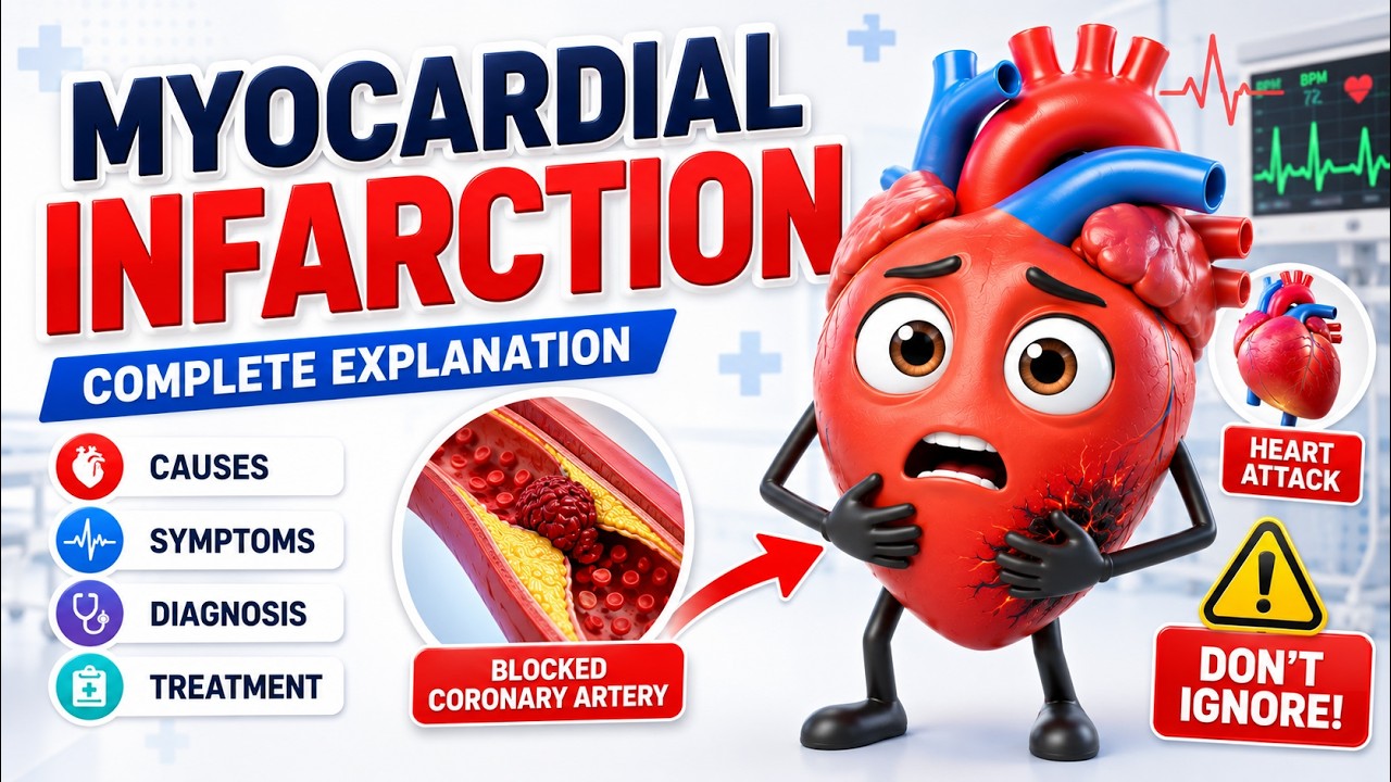
A Myocardial Infarction (MI), colloquially known to the public as a "heart attack," represents a profound, life-threatening medical emergency and remains one of the leading causes of mortality and morbidity worldwide. It is the most severe manifestation of Acute Coronary Syndrome (ACS). At its core, an MI occurs when the essential flow of oxygen-rich blood to a specific section of the heart muscle (the myocardium) is abruptly and severely restricted or entirely completely obstructed. The human heart, a relentless pump, relies on a continuous, uninterrupted supply of oxygen and metabolic nutrients delivered exclusively through the delicate network of the coronary arteries. When a blockage occurs, typically precipitated by the sudden rupture of a fragile atherosclerotic plaque followed by rapid thrombus (blood clot) formation, the cardiac tissue immediately distal to the blockage becomes acutely ischemic. If robust blood flow is not restored rapidly through medical or surgical intervention, the heart muscle cells cross a threshold from reversible injury into irreversible necrosis (tissue death). This pathological progression leads to the classic, terrifying symptoms of a heart attack, including crushing, heavy chest pain, profound shortness of breath, severe diaphoresis (sweating), and classic referred pain radiating to the left arm, jaw, neck, or interscapular region of the back.
The Deep Pathophysiology: How a Heart Attack Actually Happens
The underlying architectural cause of the vast majority of myocardial infarctions is a chronic, progressive condition known as coronary artery disease (CAD) or coronary atherosclerosis. Over years, or often decades, a combination of circulating cholesterol (specifically LDL), oxidized fats, calcium deposits, and inflammatory macrophages build up inside the intimal layer of the coronary artery walls. This buildup forms what are known as atherosclerotic plaques. While these plaques narrow the artery and can cause stable angina (chest pain on exertion), the true danger lies in plaque instability.
A heart attack typically occurs not simply because a plaque slowly grows to block the whole artery, but rather because a vulnerable, lipid-rich plaque suddenly tears or ruptures. The body's intricate immune and hemostatic (clotting) systems immediately detect the exposed subendothelial tissue as a severe vascular injury. Platelets rush to the site, adhere to the ruptured plaque, and begin to rapidly aggregate. The coagulation cascade is aggressively activated, leading to the formation of a fibrin mesh that solidifies into a massive blood clot (thrombus). This thrombus can completely occlude the arterial lumen within a matter of minutes.
Within seconds of a total occlusion, the affected cardiac myocytes are starved of oxygen and are forced to switch from highly efficient aerobic metabolism to highly inefficient anaerobic glycolysis. This results in a rapid accumulation of toxic lactic acid, lowering the intracellular pH. The lack of ATP causes the sodium-potassium pumps to fail, leading to cellular swelling and massive shifts in resting membrane potentials (which causes the deadly arrhythmias commonly seen in acute MI). Within 20 to 40 minutes of severe, sustained ischemia, the first wave of irreversible cellular damage and necrosis begins in the subendocardium (the deepest layer of the heart muscle). Over the next several hours, a "wavefront of necrosis" progresses outward toward the epicardium. This rapid, irreversible destruction of irreplaceable cardiac tissue is the origin of the critical cardiology mantra: "Time is muscle." Every minute of delay directly correlates to permanent loss of ventricular pumping function.
STEMI vs. NSTEMI: Understanding the Critical Diagnostic Differences
When a patient presents to the emergency department, urgent care clinic, or EMS with symptoms highly suspicious of Acute Coronary Syndrome (ACS), healthcare providers must act with extreme urgency. The immediate diagnostic workup relies heavily on two pillars: a rapid 12-lead Electrocardiogram (ECG/EKG) obtained within 10 minutes of arrival, and serial measurements of highly specific cardiac biomarkers (such as high-sensitivity Troponin I or T). Based on these findings, heart attacks are broadly classified into two major, clinically distinct categories that dictate entirely different treatment pathways:
1. STEMI (ST-Elevation Myocardial Infarction)
A STEMI is the most severe, destructive, and immediately urgent type of heart attack. It occurs when a major epicardial coronary artery suffers a complete, 100% transmural blockage by a massive blood clot. Because the entire thickness of the ventricular muscle wall (transmural ischemia) is acutely deprived of oxygen, the electrical repolarization of the heart is dramatically altered. This manifests on the 12-lead ECG as a classic, unmistakable abnormality known as "ST-segment elevation" in the leads looking at the damaged area of the heart (e.g., leads II, III, aVF for an inferior wall MI). A STEMI is an absolute, code-level medical emergency. The primary, overriding clinical goal is immediate reperfusion therapy to re-open the blocked artery. This is typically achieved through Percutaneous Coronary Intervention (PCI) in a specialized cardiac catheterization lab, where a cardiologist inflates a balloon and places a stent to crush the clot and keep the artery open. If a cath lab is not available within a 120-minute drive, powerful systemic thrombolytic (clot-busting) medications like Alteplase or Tenecteplase must be administered immediately. The gold-standard target is a "door-to-balloon" time of under 90 minutes.
2. NSTEMI (Non-ST-Elevation Myocardial Infarction)
An NSTEMI occurs when a coronary artery is only partially blocked, transiently occluded, or blocked but receiving some collateral blood flow. This causes severe, critical ischemia and damage to a portion of the heart muscle, but crucially, it does not typically involve the entire full thickness of the ventricular wall. Because the damage is primarily subendocardial, there is no ST-segment elevation seen on the ECG (although clinicians may observe ST depression, transient T-wave inversion, or even a completely normal ECG). However, the definitive diagnosis of an NSTEMI is confirmed through blood tests showing significantly elevated cardiac biomarkers, primarily high-sensitivity Troponin. The elevated Troponin proves definitively that irreversible heart muscle damage has occurred. While slightly less immediately catastrophic in the first few hours compared to a STEMI, an NSTEMI still carries a high risk of deterioration and requires urgent, aggressive medical management. This includes robust dual antiplatelet therapy (DAPT), systemic anticoagulation (e.g., Heparin or Lovenox), and typically a planned, early invasive cardiac catheterization within 24 to 48 hours to identify and stent the culprit lesions.
The MONA Protocol: Initial Emergency Pharmacological Management
In the high-stakes environment of emergency medicine, the initial pharmacological management for a patient experiencing acute myocardial infarction was traditionally remembered by the classic mnemonic MONA. However, it is crucial for modern nursing and medical students to understand that current AHA/ACC guidelines dictate a specific order of administration and strict criteria for usage to maximize patient safety and physiological outcomes. It is not simply given blindly:
- O - Oxygen: Historically, 100% oxygen was given to every chest pain patient. Modern evidence has proven this is dangerous. Oxygen therapy is now administered only if the patient's oxygen saturation (SpO2) falls below 90%, if they are in overt respiratory distress, or if they show signs of acute heart failure. Hyperoxia (too much oxygen in the blood) in patients with normal oxygen levels can actually cause paradox coronary vasoconstriction, increase oxidative stress, and worsen the final infarct size.
- A - Aspirin: This is arguably the single most critical, life-saving early intervention that can be given by EMS or bystanders. The patient is instructed to aggressively chew 162-325 mg of non-enteric coated aspirin immediately upon symptom onset. Aspirin is a rapid-acting, potent antiplatelet drug. It permanently inhibits the cyclooxygenase-1 (COX-1) enzyme, preventing platelets from clumping together and halting the further expansion of the deadly blood clot. Chewing the tablets ensures rapid absorption directly into the bloodstream through the buccal mucosa.
- N - Nitroglycerin: Nitroglycerin is administered sublingually (under the tongue) as a tablet or a spray. It acts as a powerful, rapid-onset systemic vasodilator. It widens the coronary arteries, marginally improving collateral blood flow to the ischemic myocardium. More importantly, it profoundly dilates peripheral veins, pooling blood in the extremities and reducing venous return to the heart (reducing preload). This drastically decreases the heart's overall workload and oxygen demand, effectively relieving ischemic chest pain. (Critical Nursing Note: Nitroglycerin is strictly contraindicated if the patient has taken phosphodiesterase inhibitors like Sildenafil/Viagra within 24-48 hours, or if they have profound hypotension, or if they are experiencing a right ventricular infarction where preload is essential for survival).
- M - Morphine: Morphine is a powerful opioid analgesic historically used for severe, persistent chest pain that is totally unrelieved by three doses of nitroglycerin. Beyond basic pain relief, morphine heavily reduces patient anxiety and decreases sympathetic nervous system (fight-or-flight) activity. This in turn reduces the heart's oxygen demand by slightly decreasing heart rate and blood pressure. However, recent guidelines suggest cautious use, as morphine can delay the absorption of crucial oral antiplatelet medications like Ticagrelor or Clopidogrel.
Long-Term Nursing Care, Rehabilitation, and Secondary Prevention
Surviving the acute phase of a myocardial infarction is only the first step in a lifelong journey. Long-term medical management and nursing care focus heavily on "secondary prevention" to aggressively avoid future, potentially fatal cardiovascular events. Patients are typically discharged on a complex, multi-drug regimen that must be strictly adhered to. This generally includes:
- Dual Antiplatelet Therapy (DAPT): Usually Aspirin combined with a P2Y12 inhibitor (like Plavix or Brilinta) for at least one year to prevent clots from forming inside newly placed stents.
- Beta-Blockers: Medications like Metoprolol slow the heart rate, lower blood pressure, and reduce myocardial oxygen demand. They are proven to prevent lethal arrhythmias and improve long-term survival post-MI.
- ACE Inhibitors or ARBs: Medications like Lisinopril prevent adverse "ventricular remodeling" (the process where the damaged heart muscle dilates and becomes weak), significantly reducing the risk of developing congestive heart failure.
- High-Intensity Statins: Medications like Atorvastatin or Rosuvastatin are prescribed regardless of the patient's baseline cholesterol. They powerfully stabilize the remaining atherosclerotic plaques, drastically reduce inflammation within the arteries, and aggressively lower LDL cholesterol.
Furthermore, intensive patient education by cardiovascular nurses is paramount. This education must focus on radical lifestyle modifications such as absolute smoking cessation, adopting a Mediterranean or heart-healthy diet, participating in regular, supervised cardiac rehabilitation programs, achieving healthy weight loss, and strict management of comorbid conditions like essential hypertension and type 2 diabetes mellitus.