
Hemostasis Explained | The Clotting Cascade & Fibrinolysis (3D Animation)
Hemostasis Explained: How the Body Stops Bleeding Hemostasis is the vital, multi-step physiological process that stops bleeding (hemorrhage) when a blood vessel i...
Medically Reviewed By
Medscope Editorial Team Verified
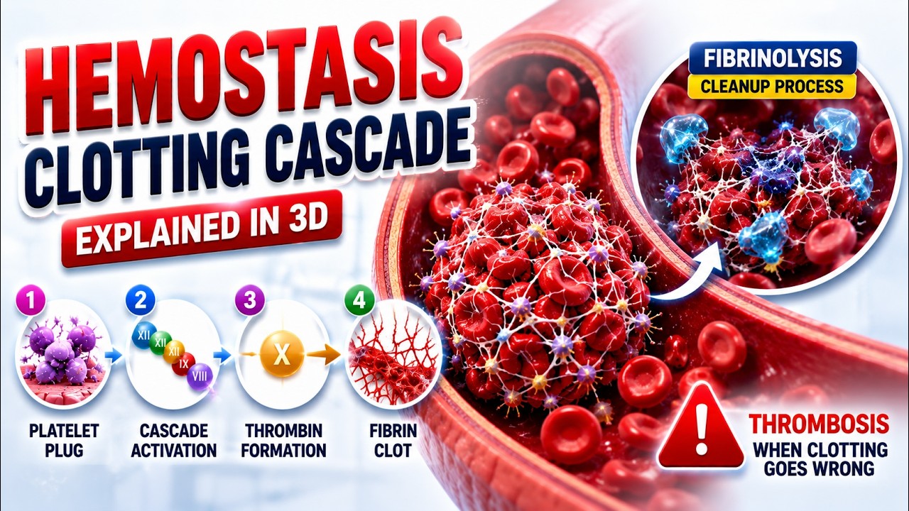
Hemostasis Explained: How the Body Stops Bleeding
Hemostasis is the vital, multi-step physiological process that stops bleeding (hemorrhage) when a blood vessel is injured. It is a highly regulated, delicate balance; the body must clot blood quickly enough to prevent exsanguination, but it must not form unnecessary clots that could cause a stroke or DVT. The process of hemostasis occurs in three distinct overlapping phases.
Phase 1: Primary Hemostasis (Vascular Spasm & Platelet Plug)
The immediate response to a cut or ruptured blood vessel happens in seconds:
- Vascular Spasm: The smooth muscle of the damaged blood vessel immediately constricts (vasoconstriction) to reduce blood flow to the injured area.
- Platelet Plug Formation: The injury exposes underlying collagen and a protein called von Willebrand factor (vWF). Platelets flowing in the blood stick to this exposed tissue, change their shape, and release chemicals (like ADP and Thromboxane A2) to call more platelets to the site, forming a temporary, soft "plug."
Phase 2: Secondary Hemostasis (The Clotting Cascade)
The temporary platelet plug is weak and can easily be washed away. To make it permanent, the body initiates the Coagulation (Clotting) Cascade. This is a complex chain reaction of proteins (clotting factors produced by the liver) activating each other.
The cascade has two starting pathways: the Intrinsic pathway (triggered by internal blood trauma) and the Extrinsic pathway (triggered by external tissue damage). Both pathways meet at a common junction (Factor X activation).
The Final Result: The cascade's ultimate goal is to convert Prothrombin into Thrombin. Thrombin is the master enzyme that takes soluble Fibrinogen and converts it into insoluble Fibrin strands. These tough fibrin strands wrap around the platelet plug like a reinforced net, creating a hard, stable blood clot.
Phase 3: Fibrinolysis (Clot Breakdown)
Once the injured tissue has fully healed, the permanent clot is no longer needed. The body releases an enzyme called Plasmin, which slowly digests and dissolves the fibrin net, clearing the blood vessel and restoring normal, smooth blood flow. This process of dismantling the clot is known as fibrinolysis.
Clinical Tests Related to Hemostasis
Healthcare professionals assess hemostasis using several common laboratory tests. Platelet count helps evaluate the primary hemostasis system. PT/INR mainly assesses the extrinsic and common coagulation pathways and is used to monitor warfarin therapy. aPTT evaluates the intrinsic and common pathways and is commonly monitored during unfractionated heparin therapy. Fibrinogen and D-dimer levels may be checked when disseminated intravascular coagulation, thrombosis, or abnormal clot breakdown is suspected.
Why Hemostasis Balance Is So Important
If hemostasis is too weak, the patient may develop easy bruising, prolonged bleeding, nosebleeds, heavy menstrual bleeding, internal bleeding, or dangerous postoperative hemorrhage. If hemostasis is too strong, unnecessary clots can form inside vessels, causing deep vein thrombosis, pulmonary embolism, heart attack, or stroke. This is why anticoagulants and antiplatelet drugs must be monitored carefully. The body is constantly balancing bleeding risk against clotting risk, and even small changes in platelets, clotting factors, liver function, vitamin K, or medications can shift that balance.
Clinically, any unexplained bleeding or clotting event should prompt a focused review of medications, liver function, platelet count, coagulation tests, family history, and recent illness.